Previous Winners
ARTiS has been around since 2013, it started of as an internal competition but has since grown and made a leap to open to the public in 2015. With the competition in 2016 sponsors like Lundbeckfonden have entered and started to support the contest, this allowed to increase the reach significantly and people from all over the world have since participated. The winning pictures of the previous years can be found on this page.
The winning pictures from the ARTiS 2019 Competition.
Best ARTiS Image & Category: Mini - A demonstration of diffusion
by Lasse Saaby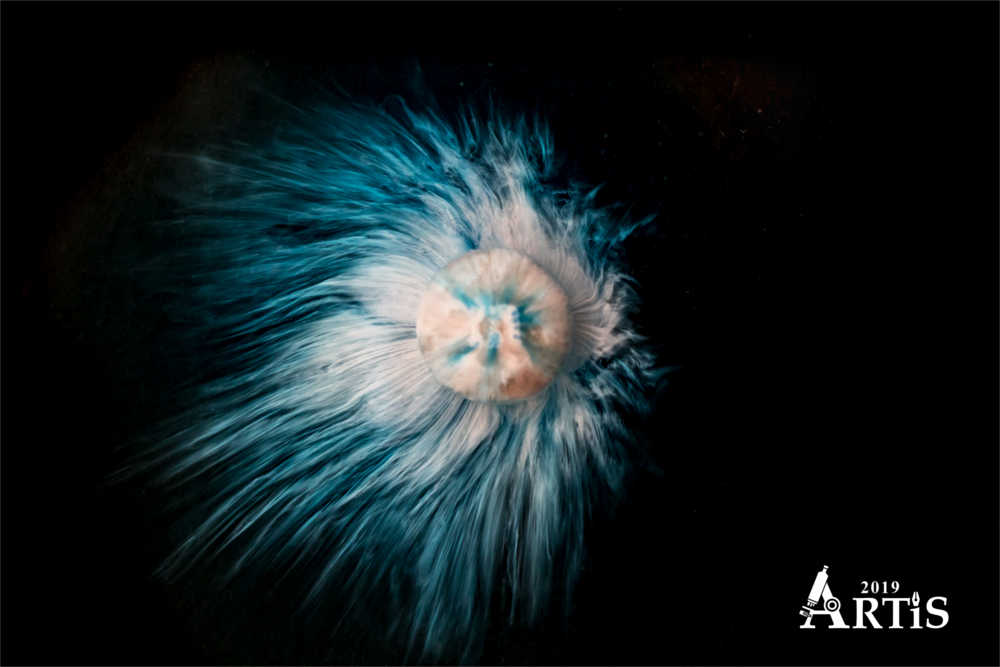
In this picture a blue M&M was placed in a petri dish filled with purified water. The outer layer of blue food dye dissolves almost immediately. Subsequently, the underlying white sugar shell starts to dissolve and falls to the bottom of the petri dish around the M&M as partially dissolved sugar. The stripes moving away from the M&M are created by movement of dissolved sugar through the layer partially dissolved sugar and blue food dye. As the sugar around the M&M dissolves it will move outward away from the M&M, where the sugar concentration is high, to the surrounding water where the sugar concentration is low - this is called diffusion.
Category: Macro - Silent Dance
by Neža Cankar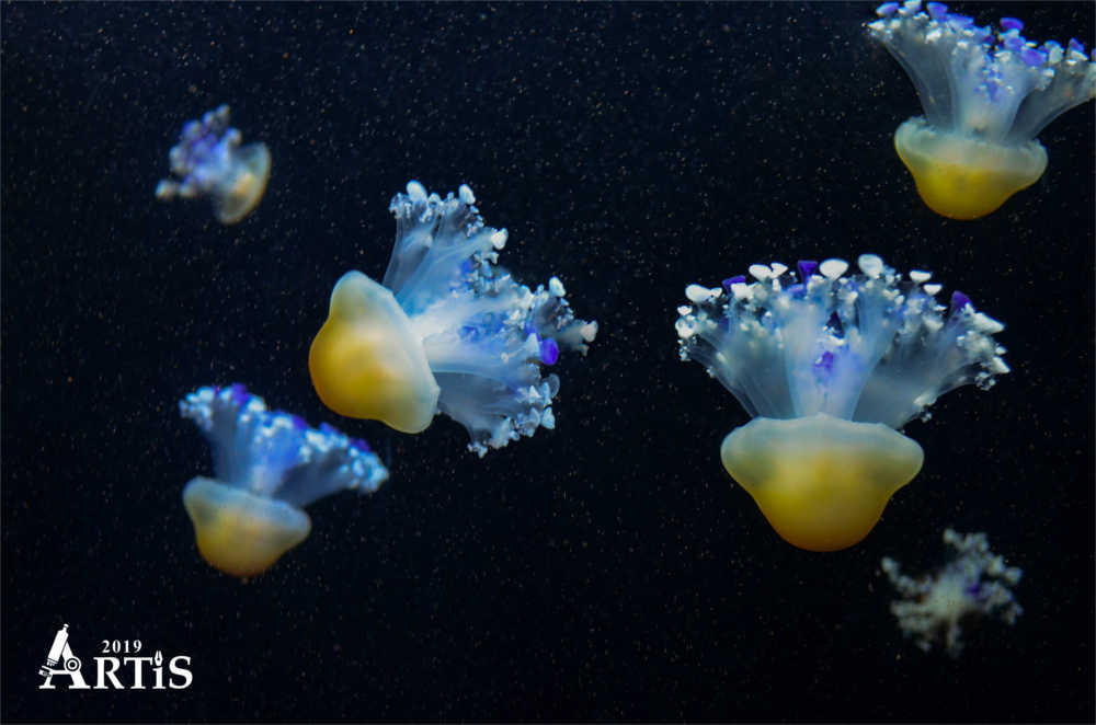
Jellyfish are one of those astonishing but intimidating aquatic animals. They already existed millions of years before dinosaurs. Near 99% of their body mass is water and yet they look extremely elegant. Furthermore, they had greatly revolutionized scientific research after the green fluorescent protein (GFP) was discovered in one of the jellyfish species. GFP became tremendously useful tool in modern science and medicine as it opened a new perspective - to look directly into inner world of cellular activity.
Category: Micro - Brain tumor in a jar
by Liselotte Jauffred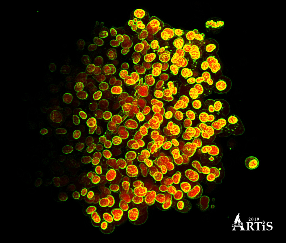
Brain tumor in a jar: A small lump of cancer cells that we have grown to resemble a small spherical tumor (1/10 mm). The nuclei of the cells are colored red and the outermost green rings show the surface membranes. We recorded this with the so-called light-sheet microscopy technique that allows us to image the cells in the tumor, even the innermost central part. We can therefore use this technique to make 3D models of brain tumor growth and determine how the geometric location of the individual cells affects their shape and ability to invade the surrounding brain tissue (Kristoffer Laugesen & Liselotte Jauffred, Niels Bohr Institute)
Category: Nano - Herpes Simplex Virus 2
by Victor Padilla-Sanchez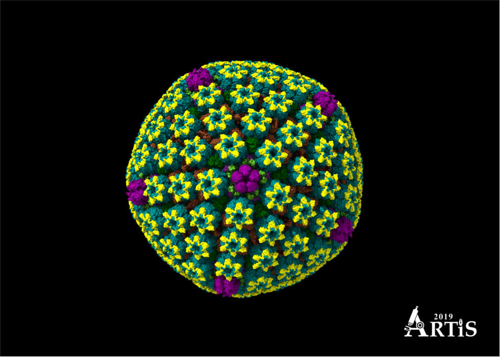
Herpes simplex virus 2 at atomic resolution of 3.1 Angstroms. This capsid has recently been resolved by cryoEM reconstruction in 2018 having a diameter of 125 nm and being composed of approximately 3000 different proteins. HSV2 causes genital ulcer disease as a sexually transmitted virus. This image has been constructed in ChimeraX from University of California San Francisco in a high-performance computer.
Category: Visualizations - High Energy Particles Collision
by Girolamo Sferrazza Papa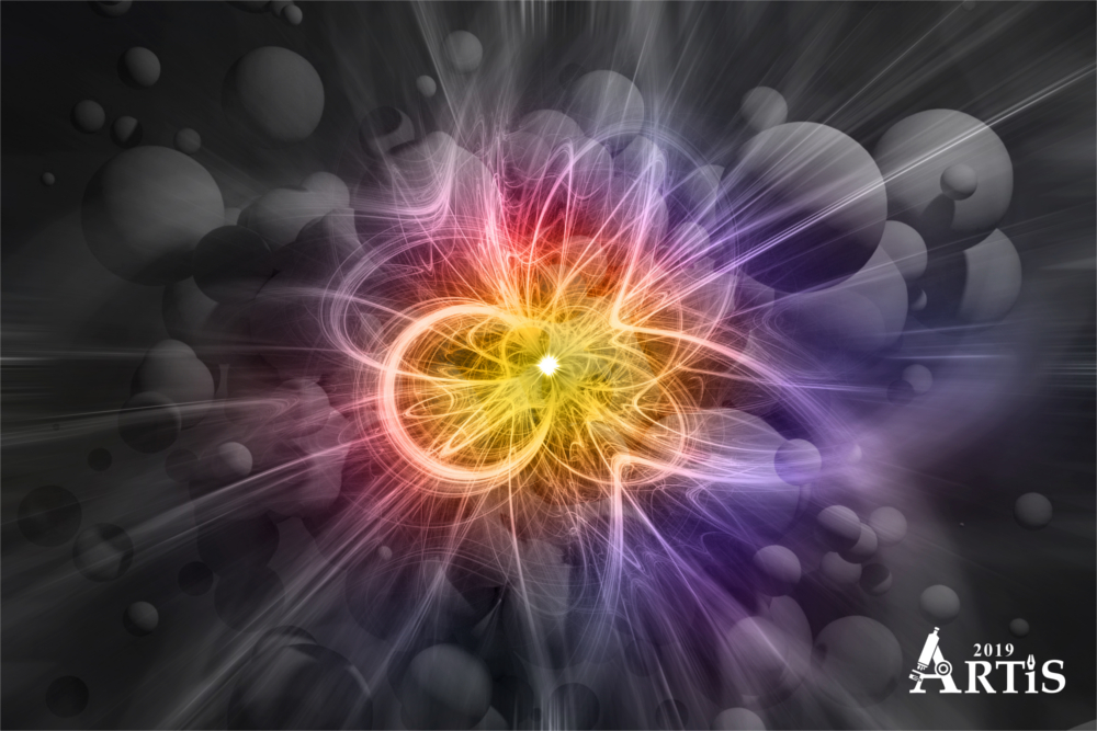
Abstract representation of high energy subatomic particles collision.
Category: ARTiS Young - Farvet ild
by Victor Haargaard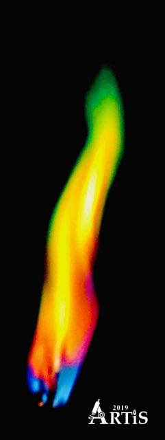
I find fire fascinating and beautiful, yet it can be so destructible. I have taken a picture of a flame I have colored with a mixture of Calcium sulphate, CaSO4, Copper sulphate, CuSO4, and table salt, NaCl. The reason why the flame gets this color is because of the emission spectrum. A molecule or an atom jumps from a higher energy level to a lower energy level in a few nanoseconds or less. While the electron from an photon relaxes to a lower state, magnetic radiation is released resulting in different colors depending on which element are used.
Category: Humouristic category - Kiss please
by Viktor Sykora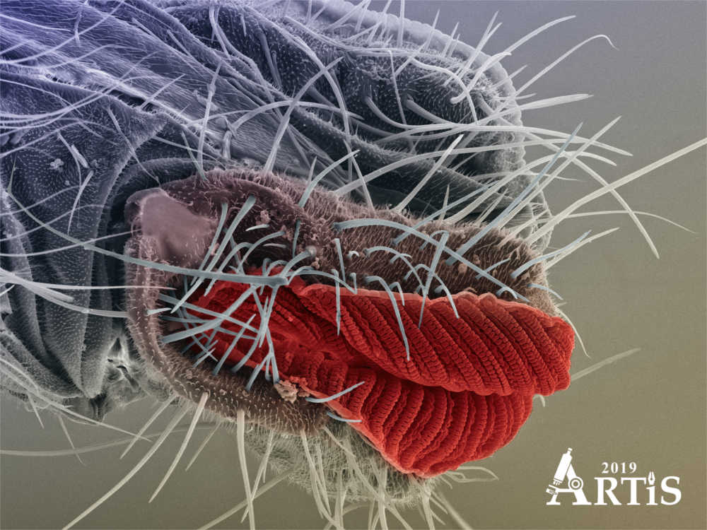
Proboscis is part of the mouth of the Houseflies, which serves to tasting and eating food. The end part is called Labellum.
Category: Best instrument - Where to next?
by Mikkel Roald-Arbøl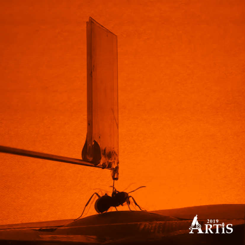
What goes on in the mind of an ant? A neuroscientific approach to this question is recording neural activity in a freely behaving animal; it is a highly desirable goal but enormously tricky to do in an animal of only 2 cm. This picture is taken the very first time a wood ant engages with and navigates within a virtual environment. As the ant walks on top of a styrofoam ball, it moves around in a custom-made environment – just like in a computer game. My name is Mikkel Roald-Arbøl and this is the culmination of my master research in the ‘Laboratory of Evolutionary Computational Neuroscience’ at University of Sussex with Dr. Jeremy Niven. The photo is a still frame from a normal video recording, with an additional frame superimposed to suggest the movement of the ant. Original colours are BW.
Culture Night public choice - Brainheart
by Søren Heide Jørgensen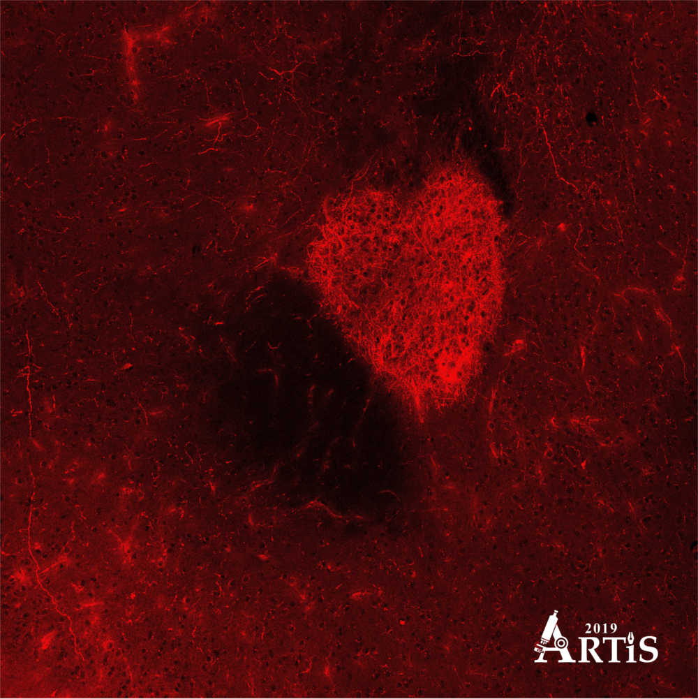
This heart-shaped brain structure is only 0,4 millimeters high and 0,35 millimeters across and is comprised of thousands of tiny entangled brain fibers containing the signaling molecule dopamine. The fibers come from dopaminergic brain cells located far away, and end here in the region called Nucleus Accumbens, where dopamine is released when something pleasurable or important happens. In my research, I am investigating exactly when dopamine is released during different behaviors, and it just so happens, that when a male mouse sees or sniffs a female mouse a large amount of dopamine is released – I like to think that the brainheart is flashing.
Category:G protein-coupled receptors - Wrapped in fat
by Alexander Hauser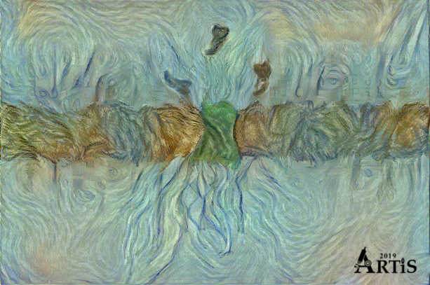
For many years, people realized that cells had to be able to signal information to each other in some way. But how? There had to be some kind of entity that would transmit the signal (such as hormones shown on top) from the outside of the cell membrane barrier somehow to set off a response inside the cell. These entities are receptors (green in centre) like G-protein coupled receptors (GPCRs) embedded in the fatty cell membrane or lipid bilayer. We are studying GPCRs through molecular simulations and evolutionary biology in order to understand the human hormone signaling system and to leverage their accessibility through targeted drug design. The image is an artistic representation through a neural network-based art approach from a previously rendered atomic representation of the cannabinoid receptor obtained by X-ray crystallography.
The winning pictures from the ARTiS 2018 Competition.
Best Image & Category: Micro - Protecting the pancreas
by Kristian Jensen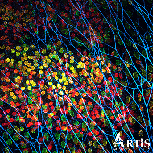
The pancreas is protected by a transparent net of cells. The protective layer is thinner than a hair width and here coloured blue, while the core of the cells beneath is rainbow-coloured.
Humoristic - The Adventure of 'Calagon'
by Cheng Choo Lee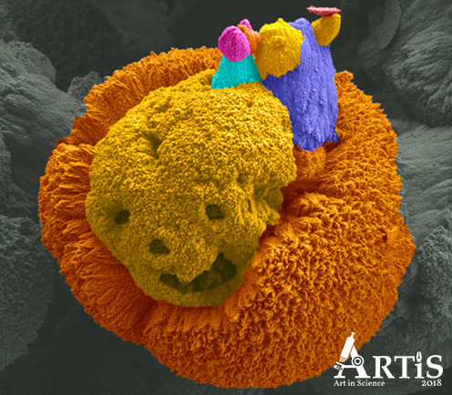
We are looking at cryo-FESEM image of a mineral comprised of calcite (particle agglomerate) and aragonite (rod-like shape). Interestingly, the captured image also showed a pair of creatures (a stone-aged man with his piggy pet) on the 'mineral' that looks like a 'skeleton-skull'. The image was taken and colorized by Cheng Choo Lee (Umeå Core Facility for Electron Microscopy, UmU) and the sample was courtesy of Elin Tollefsen (Geological Sciences, SU).
Culture Night Visitors Choice - Pathways in the human brain (2)
by Henrik Groenholt Jensen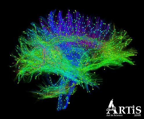
Diffusion-weighted MRI is used to non-invasively trace neuronal pathways by following the flow of hydrogen protons as they diffuse within and around cells. Here, we see a side-view of pathways in the brain, coloured by the orientation of the fibers. One such is the blue pathways moving up from the brain stem and radiating out. By following this movement of water, we get an indication of how the brain is "wired" through the white matter fiber tracts connecting the "computational units" of the grey matter neurons. My name is Henrik G. Jensen and a recent PhD graduate from Computer Science. I have been working on the complex task of comparing diffusion-weighted images by creating spatial maps between scans, profiling groups and the temporal and/or pathological evolution in single subjects.
Facebook Public Choice & Category: Visualisations - Snowflake
by Behbood Abedi ft. Bruno Fonseca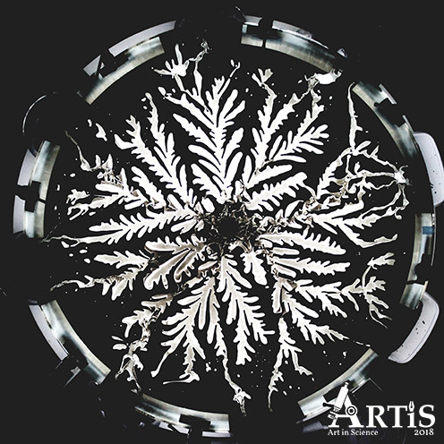
This is the Hele-Shaw flow between two parallel flat plates separated by an infinitesimally small gap. A fluid is injected into the shallow geometry from below where it is bounded by another liquid or gas. Various problems in fluid mechanics can be approximated to these flows; so the study of them is of great importance. In this case: air penetrates a viscous polymer with high injection rate, so the viscous fingering generates this gorgeous snowflake.
Category: Nano - Nano Vineyard
by Wonjong Kim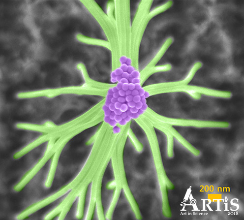
Hello, my name is Wonjong Kim. I am a second year PhD student in EPFL, Switzerland. Currently, I am working on nanowire array based solar cells. Here is an SEM image of GaAs nanowires on a silicon wafer. You can see nanoscale grapes cultivated in the clean room facilities. Very uniform and well-ordered nanowires are touching each other and made a beautiful network after a reactive ion etching process.
Category: Mini - Pathways in the human brain
by Henrik Groenholt Jensen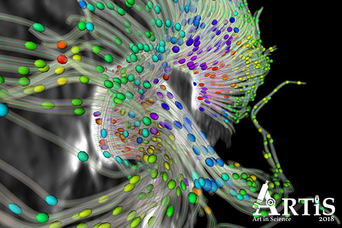
Diffusion-weighted MRI is used to non-invasively trace neuronal pathways by following the flow of hydrogen protons as they diffuse within and around cells. Here, we see a cross-section of the brain bridge, the corpus callosum, connecting its two hemispheres. The coloured glyphs indicate the measured direction of the diffusion along fiber pathways. By following this movement of water, we get an indication of how the brain is "wired" through the white matter fiber tracts connecting the "computational units" of the grey matter neurons. My name is Henrik G. Jensen and a recent PhD graduate from Computer Science. I have been working on the complex task of comparing diffusion-weighted images by creating spatial maps between scans, profiling groups and the temporal and/or pathological evolution in single subjects.
Category: Macro - Swords Fighting
by Jonas Drotner Mouritsen and Ole G. Mouritsen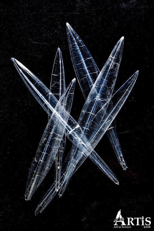
Squid-gladius-0487.jpg ‘Swords fighting’ The gladiuses (chitin backbones) of squids (Loligo forbesii) photographed on a black and reflecting surface of a stove. Scale: Max 2 cm Technique: ordinary photography with digital camera. Research relation: Investigation of the gastrophysics of Danish squid. Contributers: photographer: Jonas Drotner Mouritsen, Chromascope; scientist: professor Ole G. Mouritsen, Department of Food Science, University of Copenhagen.English Cephalopods, specifically Coleoidea (squid, octopus, and cuttlefish), have for millennia been used as marine food by humans across the world and across different food cultures. It is particularly the mantle, the arms, the ink, and part of the intestines such as the liver that have been used. In addition to being consumed in the fresh and raw states, the various world cuisines have prepared cephalopods by a wide range of culinary techniques, such as boiling and steaming, frying, grilling, marinating, smoking, drying, and fermenting. Cephalopods are generally good nutritional sources of proteins, minerals, omega-3 fatty acids, as well as micronutrients, and their fat content is low. Whereas being part of the common fare in, e.g., Southeast Asia and Southern Europe, cephalopods are seldom used in regional cuisines in, e.g., North America and Northern Europe although the local waters there often have abundant sources of specific species that are edible. There is, however, an increasing interest among chefs and gastroscientists to source local waters in a more diverse and sustainably fashion, including novel uses of cephalopods to counterbalance the dwindling fisheries of bonefish, and to identify new protein sources to replace meat from land-animal production. Combining these trends in gastronomic development with the observation that the global populations of cephalopods are on the rise holds an interesting promise for the future. There are three main groups of cephalopods: octopus, torpedo-shaped, and sepia-like, and they have only few hard part in their body. The octopus has a hard beak made of chitin, the torpedo-shaped has additionally a so-called sword (gladius) made of chitin, and the sepia-like has a beak of chitin and a calcareous backbone (cuttlebone). As part of a multidisciplinary research project on the gastrophysics and culinary applications of cephalopods, a number of photographic images were made of both the hard and soft parts of Danish squid.
The winning pictures from the ARTiS 2017 Competition.
1st prize, Culture Night, Category: Mini - Beauty in Seawater
by Jannicke Wiik-Nielsen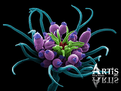
Coloured scanning electron micrograph (SEM) of the hydroid, Echtopleura larynx. The hydroid is a fouling organism usually found attached to sunken ropes, floating buoys, mussel shells, rocks and seaweed. The hydroid has two distinct rings of tentacles, one around its mouth and the other at the base of the head. In between the two rings of tentacles, are the gonophores, or the sexual buds. The hydroids are beautiful but beware; their delicate looks belie their potent nature. They possess an armament of stinging cells equipped with nematocysts in their tentacles used to capture and subdue prey. Magnification: x20 when printed at 10cm wide.
I'm a researcher at the Norwegian Veterinary Institute. The institute uses my images in different communication contexts and supplies the images to Science Photo Library.
Instrument, Social Media, Category: Macro - Precious bequer in flames
by Helena Augusta Lisboa de Oliveira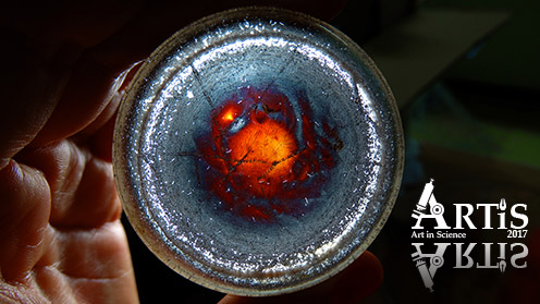
This is a beaker containing nanoparticles.
Humor, Category: Nano - Agony
by Fabrizio Gualandris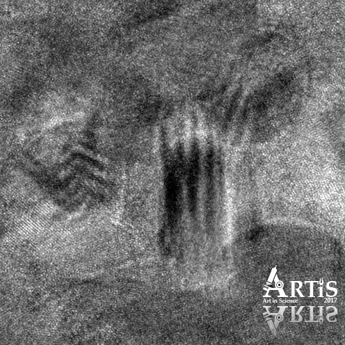
The image was taken using a transmission electron microscope looking to a ceramic sample. each doats is an atom! it represents the agony that each PhD student, at least time, ever felt in his/her job.
Category: Micro - Erotic Art
by Jannicke Wiik-Nielsen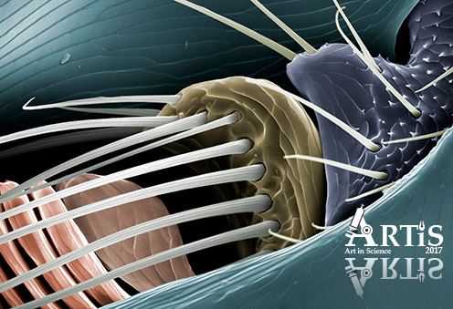
Coloured scanning electron micrograph (SEM) of a dog flea antenna. The fleas particularly live on domestic dogs and cats, but may occasionally bite humans. The fleas have antennae on both sides of their head. These are delicate sense organs which play a significant role in host-finding and are crucial for successful mating. They contain disc-shaped clasping organs which secretes an adhesive, glue-like substance. With erect antennae, the male flea moves towards a female until their heads contact each other. He then uses his antenna to push the female’s abdomen up until he is underneath her and then uses his claws to grab onto the female’s legs during mating. Magnification: x950 when printed at 10cm wide.
I'm a researcher at the Norwegian Veterinary Institute. The institute uses my images in different communication contexts and supplies the images to Science Photo Library.
Category: Mega - Fail
by Carlos Gómez Guijarro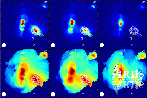
This composition of pictures displays a merger of galaxies 100,000 light years across just 1Gyr after the Big Bang. The images were taken with the Hubble Space Telescope (credit: HST/ALMA projects 2109/2012.1.00978.S, P.I.: A. Karim) using three different filters (columns), color coded and treated aiming at displaying the faintest features of the object (rows). While the result looks appealing to the eye, it is not a fair representation of the system. It is instead the product of a wrong application of the method, reflecting the learning process of the PhD student. Quoting Alberto Giacometti: “The object of art is not to reproduce reality, but to create a reality of the same intensity.” Although in this case, creation came from an unrepeatable accident in an attempt of representing reality.
Author: Carlos Gómez Guijarro, PhD student (Dark Cosmology Centre, NBI). Studying the evolutionary link between primitive starburst and present-day massive galaxies.
Category: Visualizations - Finding hay in a needle stack
by Michael Asger Andersen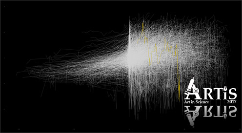
Chronic lymphocytic leukemia is a malignancy of the mature B-cells. It is the most common leukemia in the Western world, and there are an ever increasing number of new patients diagnosed in Denmark every year, with 550 patients diagnosed in 2015, compared to 300 patients in 2010. The heterogeneous disease course is a hallmark of CLL. The median survival can be under 3 years in high-risk patients and over 25 years in low-risk patients. Our group seeks to discover patterns predictive of the disease course.
The illustration shows the evolution of the lymphocytes over time for 4000 CLL patients. The x-axis is the number of years since diagnosis, and the y-axis is the lymphocyte count in a log10 scale. The yellow line shows the lymphocyte count for a single patient.
Best scientific explanation - Malet i sten
by Magnus August Ravn Harding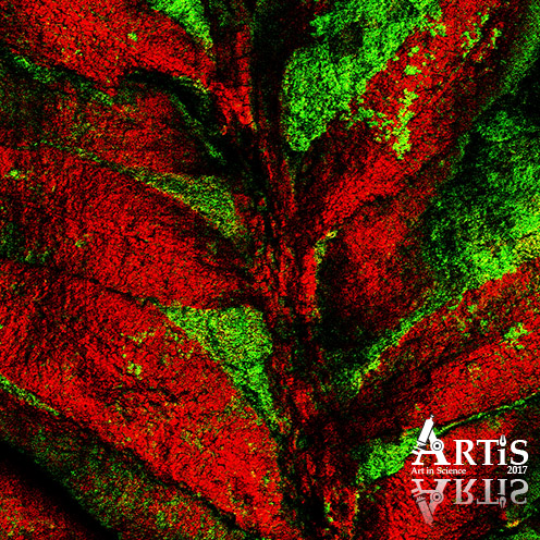
Dette billede viser fordelingen af kalk (rød) og ler (grøn) i et fossil fra samlingerne på Geologisk Museum. Billedet fortæller en omtrent 60 millioner år gammel historie fra det vestlige Grønland. Dengang voksede hér både løv- og nåletræer, herunder blandt andet denne Taxodium. Billedet vidner om en detaljeret kalkkrystallisation af hver eneste celle i det ældgamle blad og viser hvordan organismer kan bevares i sten gennem millioner af år. Således kan for længst uddøde dyr og planter samt deres levesteder efterfølgende studeres.
Billedet blev taget med time-of-flight secondary-ion-mass-spectrometry (TOF SIMS). Fossilets overflade blev bombarderet af Bismuth-ioner, som frigjorde atomer fra de øverste få nanometer af fossilet, som dernæst kunne identificeres. Hele billedet måler 5x5 millimeter.
Mit navn er Magnus Harding. Kion Norrman fra DTU Risø hjalp mig med at tage billedet til brug i mit bachelorprojekt, som jeg skriver under vejledning af Tais W. Dahl fra SNM.
Amateur Prize - Only the lonely
by Carsten Wraae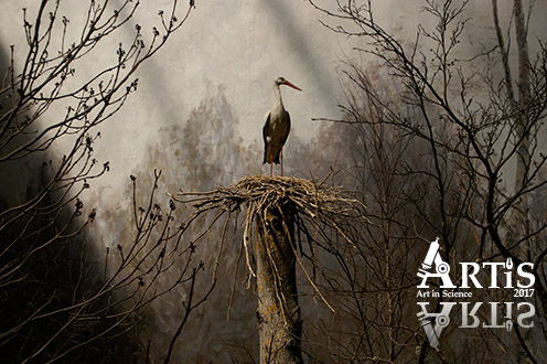
Capturing wildlife can be tricky. Unless it's "still wildlife"... The picture is taken at 'Biologiska Museet' in Stockholm, a wonderful (if slightly dusty) place where dioramas of wildlife are only lit by natural light. The lasting moment caught here is of a 'ciconia ciconia' or 'white stork' standing proudly in its nest - maybe waiting for its mate?
Young art-scientists - Flower
by Andrii Lapytskyi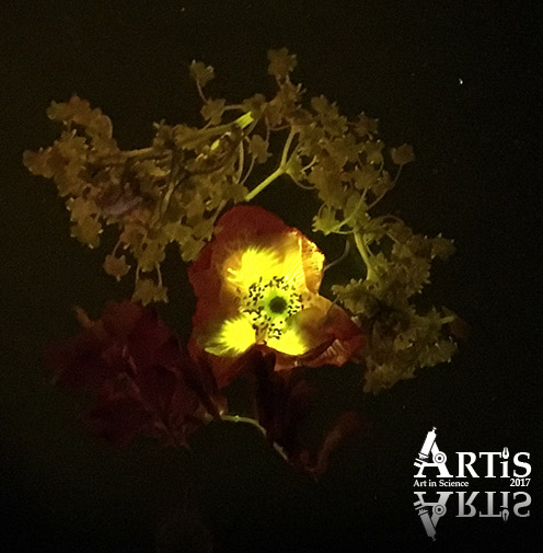
Fluorescent flowers seen through the filters.
The winning pictures from the ARTiS 2016 Competition.
1st prize - The Kiss
by Zoe Anderson-Jenkins from University of Copenhagen. The prize was a professional photo session and printed picture.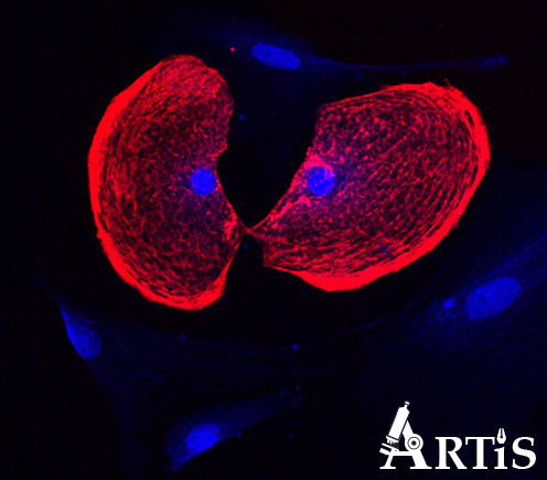
2nd prize - The Lonesome Nomad
by Benoît Desbiolles & Valentin Flauraud from EPFL in Switzerland. The prize was printed picture, Artis cup and a voucher.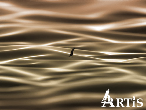
3rd prize - Form, Function, Fabulous
by Kim N. Dalby from University of Copenhagen. The prize was printed picture, Artis cup and champagne tasting.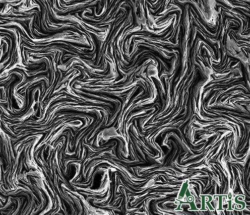
Runner-ups for best picture
Nematic Droplets
by Vance Williams from Simon Fraser University. The prize was an Artis cup.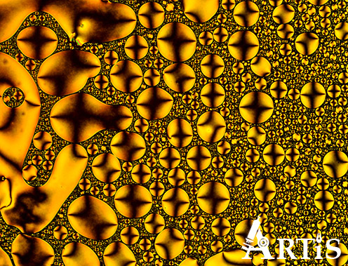
Basidiomycota
by Sebastian-Alexander Stamatis from University of Copenhagen. The prize was an Artis cup.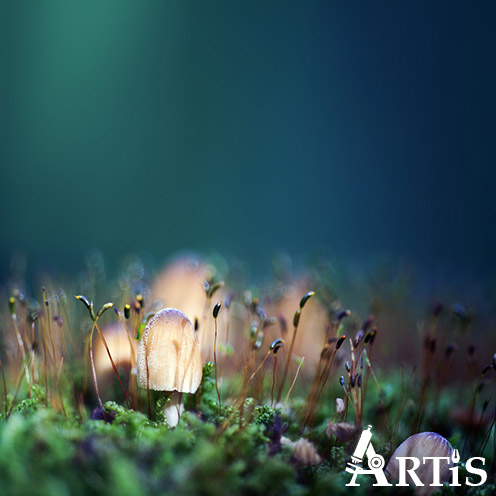
The Beginning
by artist Jens-Ole Bock (Kongstadbock) from Denmark. The prize was an Artis cup.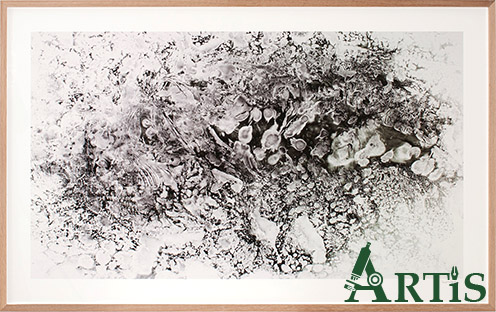
Young ART-Scientists prize
Surface Tension
by Alix and Sacha Guilbert, Morvarid, Stefan and Vilhelm from Den Dansk-Franske Skole in Copenhagen. The prize was printed picture, a visit to our lab and a trophy.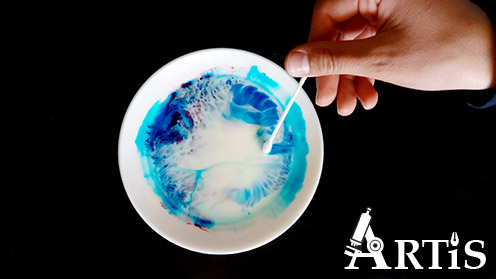
Amateur prize
Spider Web Diffraction
by Svend Erik Westh Hansen from Nature Photographers in Denmark. The prize was printed picture.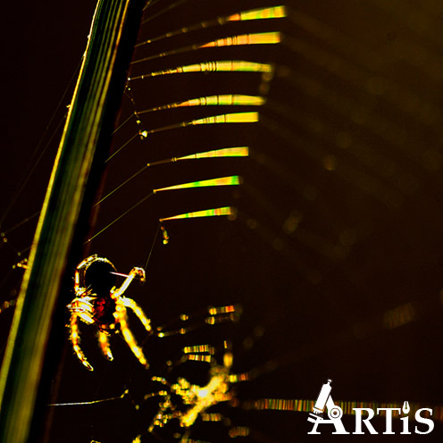
Humoristic prize
Insect samples are also good for a laugh
by Malene Fogh Bang from University of Copenhagen. The prize was a gift.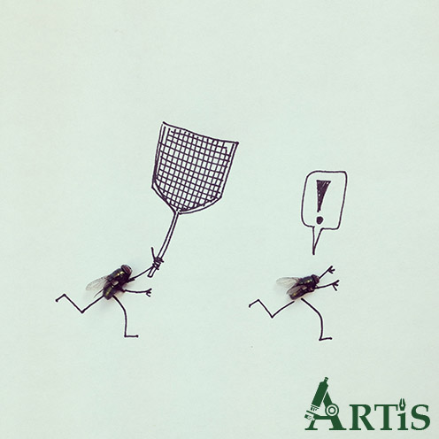
Culture Night Visitors prize
Basidiomycota
by Sebastian-Alexander Stamatis from University of Copenhagen. The prize was a gift.
Facebook prize
Magnetic Mandala
by Helena Augusta Lisboa de Oliveira from University of Brasília. The prize was a gift.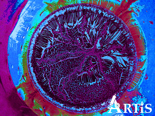
In 2015 there was no formal award ceremony, nonetheless there have been a few pictures standing out.
Dots
Forest
Strokes
Chip Carrier
Microscope 2
Heart
Microscope
